
RNase, short for Ribonuclease, is an enzyme that cleaves the phosphodiester bonds in RNA molecules, effectively breaking down RNA into smaller fragments. It is commonly known as an RNA-digesting enzyme and plays a vital role in RNA metabolism, gene regulation, and cellular defense.
RNase is a classic example of a protein with a well-defined tertiary structure, making it a key subject in biochemistry and molecular biology courses. Understanding what RNase does — from RNA degradation to immune defense — is fundamental to both academic study and modern biotechnology research.

Key basic facts about RNase:
- The molecular weight of RNase is 13,700 Da
- Christian Anfinsen (1950) and William Stein (1958) elucidated the complete structure of bovine pancreatic RNase
- RNase consists of 124 amino acids in a single polypeptide chain with 4 disulfide linkages
- RNase is hydrolyzed by the enzymes Pepsin and trypsin.
- RNase is an enzyme that cleaves the P–O (phosphodiester) bonds in RNA, which is a commonly searched fact in biochemistry
What Does RNase Do?
The primary function of RNase is to catalyze the hydrolysis of RNA—meaning it breaks the backbone of RNA molecules by cleaving phosphodiester bonds. This seemingly simple action has far-reaching biological consequences.
Here is what RNase does in living cells:
- RNA turnover: All organisms continuously produce and degrade RNA. RNase enzymes ensure that cellular RNA that is no longer needed is efficiently cleared, preventing toxic RNA accumulation.
- RNA maturation: RNases are essential for processing precursor RNA molecules into their mature, functional forms—including messenger RNAs (mRNAs), transfer RNAs (tRNAs), and ribosomal RNAs (rRNAs).
- Gene regulation: By controlling the stability and lifespan of mRNA, RNases directly regulate which proteins get made and in what quantities—a core mechanism of gene expression control.
- Antiviral defense: RNA viruses replicate using RNA as their genetic material. Active RNA degradation by RNase enzymes serves as a first line of defense against RNA virus infections. This also provides the molecular foundation for advanced cellular immune strategies like RNA interference (RNAi).
- RNase enzyme in research: Because RNase is one of the hardiest and most well-characterized enzymes in biochemistry, it is widely used as a molecular tool in laboratory research, including RNA sequencing, structural biology, and gene expression studies.
RNase Full Form and Classification
RNase’s full form is Ribonuclease. The term combines “ribonucleic acid” (RNA) and “-ase,” the standard suffix for enzymes. So, RNase is literally the enzyme responsible for nucleic acid (RNA) degradation.
Ribonucleases are classified into two major categories based on where they cleave the RNA chain:
- Endoribonucleases — cut RNA at internal sites within the chain
- Exoribonucleases — chew RNA from either the 3′ or 5′ end
Both classes fall under enzyme classification EC 2.7 (phosphorolytic enzymes) and EC 3.1 (hydrolytic enzymes).
Types of RNase: Major Endoribonucleases
Endoribonucleases cleave RNA at internal positions. Below are the major types of RNase in this category:
1. RNase A
RNase A function is one of the most studied in biochemistry. RNase A (e.g., bovine pancreatic ribonuclease A) is sequence-specific for single-stranded RNA and cleaves at the 3′ end of unpaired cytidine (C) and uridine (U) residues. Its RNase A mechanism involves a 2′,3′-cyclic monophosphate intermediate, ultimately producing a 3′-phosphorylated product. RNase A is remarkably stable — it can survive boiling in crude cellular extracts, making it one of the most robust enzymes in common laboratory usage.
2. RNase H
RNase H cleaves the RNA strand in a DNA/RNA hybrid duplex, producing single-stranded DNA (ssDNA). It is a non-specific endonuclease that catalyzes cleavage via a hydrolytic mechanism, assisted by a divalent metal ion. RNase H leaves a 5′-phosphorylated product and is critical in DNA replication and retrovirus replication (e.g., HIV reverse transcriptase contains RNase H activity).
3. RNase I
RNase I cleaves at the 3′ end of single-stranded RNA (ssRNA) at all dinucleotide bonds, leaving a 5′-hydroxyl and 3′-phosphate via a 2′,3′-cyclic monophosphate intermediate.
4. RNase III
RNase III cleaves ribosomal RNA (16S rRNA and 23S rRNA) from transcribed polycistronic RNA operons in prokaryotes. It also processes double-stranded RNA (dsRNA)—the Dicer family of RNases belongs here. Dicer cuts pre-miRNA (60–70 bp long) at a specific site, converting it into mature miRNA (22–30 bp), which actively regulates transcription and mRNA stability.
5. RNase L
RNase L is an interferon-induced nuclease. Upon activation by the immune system during viral infection, RNase L degrades virtually all RNA within the cell, halting both viral and host RNA synthesis—a dramatic but effective antiviral strategy.
6. RNase P
RNase P is unique: it is a ribozyme — a ribonucleic acid molecule that acts as a biological catalyst, just like a protein enzyme. Its primary function is to cleave the precursor sequence from pre-tRNA molecules, producing mature tRNAs. RNase P is one of only two known multiple-turnover ribozymes found in nature. Notably, a protein-only form of RNase P (without the RNA component) has recently been discovered.
7. RNase PhyM
RNase PhyM is sequence-specific for single-stranded RNA. It cleaves at the 3′ end of unpaired adenosine (A) and uridine (U) residues.
8. RNase T1
RNase T1 is sequence-specific for single-stranded RNA. It cleaves exclusively at the 3′ end of unpaired guanosine (G) residues—widely used in RNA footprinting and sequencing experiments.
9. RNase T2
RNase T2 is sequence-specific for single-stranded RNA. It cleaves at the 3′ end of all four nucleotide residues, with a preference for adenosine (A).
10. RNase U2
RNase U2 is sequence-specific for single-stranded RNA. It cleaves at the 3′ end of unpaired adenosine (A) residues only, making it highly selective and useful for RNA structure probing.
11. RNase V1
RNase V1 is non-sequence-specific but structure-specific—it cleaves base-paired (double-stranded) nucleotide residues and is used to map RNA secondary structure.
Types of RNase: Major Exoribonucleases
Exoribonucleases digest RNA from its ends (either 3′→5′ or 5′→3′). They are essential for RNA processing, quality control, and turnover.
| Exoribonuclease | Direction | Key Function |
|---|---|---|
| Polynucleotide Phosphorylase (PNPase) | 3′→5′ | Functions as both exonuclease and nucleotidyltransferase |
| RNase PH | 3′→5′ | Exonuclease and nucleotidyltransferase; involved in tRNA 3′ end maturation |
| RNase II | 3′→5′ | Processive degradation of single-stranded RNA |
| RNase R | 3′→5′ | Homolog of RNase II; uniquely degrades RNA with secondary structures without accessory factors |
| RNase D | 3′→5′ | Processes the 3′ end of pre-tRNAs |
| RNase T | 3′→5′ | Major contributor to 3′→5′ maturation of stable RNAs |
| Oligoribonuclease | 3′→5′ | Degrades short oligonucleotides to mononucleotides |
| Exoribonuclease I | 5′→3′ | Degrades ssRNA; exists only in eukaryotes |
| Exoribonuclease II | 5′→3′ | Close homolog of Exoribonuclease I |
RNase Structure
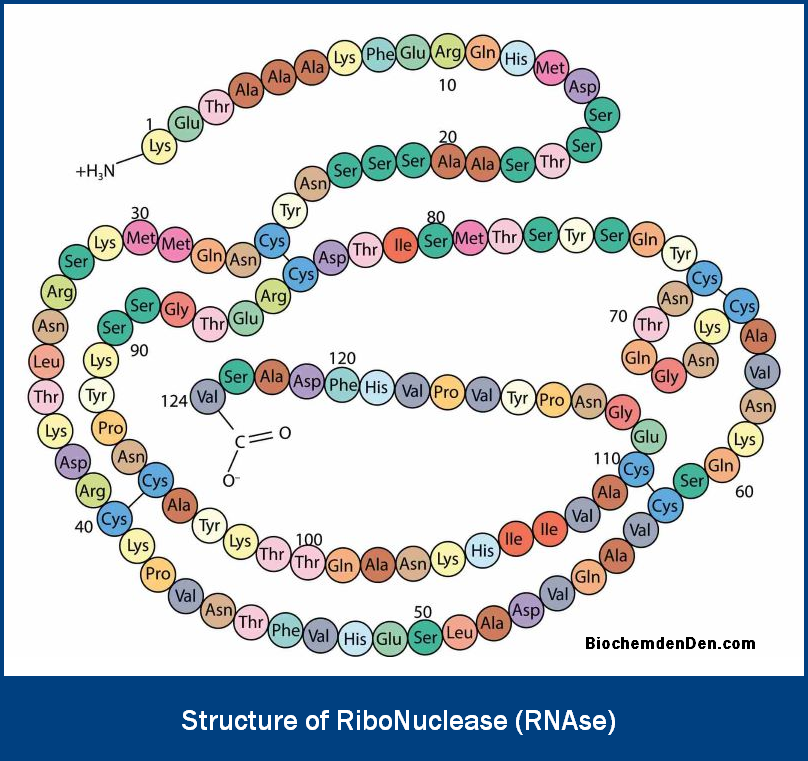
The structure of RNase A (bovine pancreatic ribonuclease A) is one of the best-characterized protein structures in biochemistry. Here are the key structural features:
- Single polypeptide chain of 124 amino acids
- Molecular weight: 13,700 Da
- Contains 4 disulfide bonds (between cysteine residues), which contribute significantly to its remarkable thermostability
- Adopts a kidney-shaped tertiary structure stabilized by hydrogen bonds, hydrophobic interactions, and the four disulfide linkages
- The active site contains two critical histidine residues (His-12 and His-119) and one lysine residue (Lys-41), which participate directly in the cleavage of the RNA phosphodiester bond
- The mechanism of RNase A follows a two-step transesterification/hydrolysis pathway: first forming a 2′,3′-cyclic phosphate intermediate, then hydrolyzing it to yield the 3′-phosphate product
- Christian Anfinsen’s landmark work on RNase A denaturation and renaturation demonstrated that a protein’s amino acid sequence alone determines its three-dimensional structure—a foundational principle of molecular biology for which Anfinsen received the 1972 Nobel Prize in Chemistry
RNase Mechanism of Action
The RNase mechanism describes how the enzyme physically breaks RNA. Using RNase A as the model:
- Substrate binding: RNase A binds single-stranded RNA, positioning a pyrimidine nucleotide (C or U) in the active site.
- Transesterification (Step 1): The 2′-OH group of the ribose attacks the adjacent phosphate group. His-12 acts as a general base (abstracting a proton), and His-119 acts as a general acid (donating a proton). This generates a 2′,3′-cyclic phosphate intermediate.
- Hydrolysis (Step 2): Water attacks the cyclic phosphate, again assisted by the two histidine residues (now switching roles), breaking the cycle and releasing a 3′-phosphate terminus and a free 5′-OH on the downstream fragment.
This two-step mechanism makes RNase A both highly specific and very efficient, explaining its widespread use in RNA biochemistry research.
RNase Enzyme Deficiency and Disease
Dysregulation or deficiency of specific RNase enzymes has been linked to several human diseases:
- RNase L deficiency has been associated with prostate cancer susceptibility and potentially chronic fatigue syndrome (ME/CFS), as RNase L plays a critical role in innate antiviral immunity.
- Mutations in RNASEH2 genes (encoding RNase H2 subunits) cause Aicardi-Goutières syndrome, a severe neurological disorder that mimics congenital viral infection.
- Elevated RNase activity in serum has been studied as a potential biomarker for certain cancers and inflammatory conditions.
- Disruption of the RNA exosome (which contains multiple exoribonucleases) is linked to various neurodegenerative diseases and hematological malignancies.
Understanding RNase function is therefore not just academically important — it has direct implications for diagnosing and treating human disease.
Importance of RNase in Living Organisms
RNases are among the most ancient and universally conserved enzymes in biology. Their presence across all domains of life — bacteria, archaea, and eukaryotes — reflects how fundamental RNA degradation is to cell survival.
- All organisms studied contain multiple RNase classes, confirming that RNA turnover is an evolutionarily ancient and indispensable process
- RNases regulate gene expression at the post-transcriptional level by controlling mRNA stability and abundance
- They are essential for the maturation of structural RNAs including rRNA and tRNA, which are required for protein synthesis
- RNA degradation by RNases forms the biochemical backbone of RNAi (RNA interference)—a powerful gene-silencing mechanism used in both natural immunity and modern gene therapy
- In biotechnology and research, RNase enzymes are indispensable tools for RNA mapping, sequencing, structural probing, and contamination removal in molecular biology labs.
Frequently Asked Questions (FAQs)
What is RNase (Ribonuclease)?
RNase, or ribonuclease, is an enzyme that breaks down RNA by cleaving its phosphodiester bonds. It is often called an RNA-digesting enzyme because it cuts RNA into smaller fragments, helping cells control RNA levels and maintain proper RNA metabolism.
What is the function of RNase in cells?
The main function of RNase is to degrade RNA molecules that are no longer needed, damaged, or still immature. By controlling RNA turnover, RNase helps regulate gene expression, supports RNA processing, and keeps the cell’s RNA pool clean and functional.
What does RNase do in simple terms?
In simple terms, RNase works like a molecular “scissor” for RNA. It recognizes RNA molecules and cuts their backbone, turning long RNA strands into smaller pieces. This prevents the buildup of excess RNA and allows cells to recycle their RNA components efficiently.
What is the full form of RNase?
The full form of RNase is ribonuclease. “Ribo” refers to ribonucleic acid (RNA), and “nuclease” refers to an enzyme that cuts nucleic acids. So, ribonuclease literally means an enzyme that specifically digests RNA.
What are the main types of RNase?
RNases are broadly divided into two groups: endoribonucleases and exoribonucleases. Endoribonucleases, such as RNase A, RNase H, and RNase III, cut RNA at internal sites. Exoribonucleases, like RNase II and RNase R, remove nucleotides one by one from the ends of RNA strands.
What is RNase A, and what does it do?
RNase A is a well-known ribonuclease that specifically cleaves single-stranded RNA after cytidine and uridine residues. It is highly stable and has been used for decades as a model enzyme to study protein structure and enzyme mechanisms in biochemistry.
What is meant by RNase structure?
RNase structure refers to the three-dimensional shape of RNase proteins, especially RNase A. RNase A has 124 amino acids and four disulfide bonds, which give it a compact and very stable structure. This well-defined shape allows it to bind RNA precisely and catalyze RNA cleavage efficiently.
How does the RNase A mechanism work?
The RNase A mechanism occurs in two main steps. First, the enzyme helps form a 2′,3′-cyclic phosphate intermediate in the RNA backbone. Second, this intermediate is hydrolyzed to produce a 3′-phosphate end on the RNA fragment. These steps make RNase A a highly efficient RNA-cleaving enzyme.
Why are RNases important in living organisms?
RNases are essential because they maintain the correct balance of RNA inside cells. They remove faulty or excess RNA, help process rRNA and tRNA, and contribute to antiviral defense. Without RNase activity, RNA would accumulate and disrupt normal cell function and gene expression.
Can problems with RNase activity lead to disease?
Abnormal RNase activity or defects in RNase-related pathways can disturb RNA processing and immune signaling. Such disruptions are linked to certain genetic and immune disorders, and they are being studied for their roles in cancer and neurological diseases in molecular medicine.
Discover more from Biochemistry Den
Subscribe to get the latest posts sent to your email.